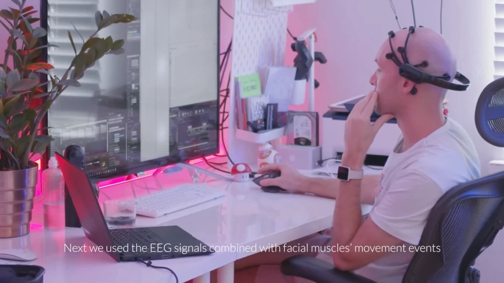
EEG Artifacts and Signal Quality: Collecting Clean, Reliable Data
H.B. Duran
Updated on
Oct 15, 2026
EEG Artifacts and Signal Quality: Collecting Clean, Reliable Data
H.B. Duran
Updated on
Oct 15, 2026
EEG Artifacts and Signal Quality: Collecting Clean, Reliable Data
H.B. Duran
Updated on
Oct 15, 2026
Introduction
Collecting EEG data is only the beginning of a successful neuroscience study. The quality of the recorded signal ultimately determines how confidently researchers can interpret brain activity and draw meaningful conclusions. Even a carefully designed experiment can produce unreliable results if the recordings are contaminated by excessive noise or physiological artifacts.
Because EEG measures electrical activity generated by the brain, it is highly sensitive to other electrical signals produced by the body and the surrounding environment. Eye blinks, facial muscle contractions, participant movement, poor electrode contact, and electrical interference can all influence the recorded signal. While many of these artifacts can be reduced during preprocessing, preventing them during data collection remains the most effective strategy for preserving data quality [1].
This guide explains the most common sources of EEG artifacts, practical techniques for reducing noise during recording, and considerations for determining when data should be corrected, rejected, or recollected.
What Is an EEG Artifact?
An EEG artifact is any recorded electrical signal that does not originate from the neural activity being studied. Artifacts can obscure genuine brain signals, reduce statistical power, and complicate data analysis if they are not recognized and managed appropriately [1].
Artifacts are a normal part of EEG research. Even experienced laboratories encounter them during data collection. The goal is not to eliminate every artifact, but to minimize avoidable sources of noise while designing experiments that produce reliable and interpretable datasets.
Researchers generally classify EEG artifacts into two broad categories: physiological artifacts generated by the participant and non-physiological artifacts introduced by equipment or the recording environment.
Physiological Artifacts
Because EEG records electrical activity from the scalp, it inevitably detects electrical signals generated elsewhere in the body. Understanding these physiological artifacts helps researchers distinguish meaningful neural activity from unrelated biological signals.
While there are measures you can take to remove some artifacts during the analysis phase minimizing artifacts during the recording is always best practice.
Eye Blinks and Eye Movements
Eye blinks produce some of the largest artifacts observed in EEG recordings because the eyes generate strong electrical potentials. These artifacts are most pronounced in frontal electrodes and can temporarily obscure nearby neural activity.
Horizontal and vertical eye movements also introduce characteristic waveform patterns that may overlap with neural responses of interest, particularly during attention and visual processing experiments.
Providing participants with opportunities to take breaks and to blink between trials and encouraging them to minimize unnecessary eye movements during critical recording periods can substantially improve signal quality [1].
Muscle Activity (EMG)
Electrical activity generated by facial muscles, jaw muscles, neck muscles, and scalp muscles often produces higher-frequency signals than typical EEG activity. Speaking, clenching the jaw, smiling, or maintaining excessive muscle tension can introduce substantial contamination into the recording.
Researchers should encourage participants to remain relaxed throughout the recording session while avoiding unnecessary facial movements whenever experimental procedures allow. Build in scheduled breaks during recordings to allow participants to relax and adjust position.
Cardiac Activity
Electrical signals generated by the heart may occasionally appear within EEG recordings, particularly depending on participant anatomy and electrode placement. Although these artifacts are generally smaller than eye movement artifacts, they can become relevant in certain experimental paradigms or longer recording sessions.
Non-Physiological Artifacts
Not all artifacts originate from the participant. Recording equipment, environmental conditions, and experimental procedures may also introduce unwanted electrical noise.
Electrode Contact
Stable electrode contact is one of the most important factors influencing EEG quality. Poor contact between the electrode and scalp increases impedance and reduces signal quality, making recordings more susceptible to environmental noise and motion-related artifacts [2].
Researchers should verify electrode placement before beginning data collection and periodically monitor signal quality during longer recording sessions. Modern EEG acquisition software often provides real-time indicators that help identify electrodes requiring adjustment.
Electrode Movement
Even when initial contact is excellent, movement of the electrodes relative to the scalp can introduce transient voltage changes that appear as artifacts.
These artifacts commonly occur when participants adjust the headset, touch their face, or make sudden head movements. Securing electrodes properly and providing clear participant instructions before recording can substantially reduce these interruptions.
Environmental Electrical Noise
EEG systems operate in environments containing numerous electrical devices capable of generating interference. Computers, monitors, lighting systems, power supplies, and nearby electronic equipment may introduce unwanted electrical signals into the recording.
Researchers should establish consistent recording environments whenever possible and minimize unnecessary electrical interference before data collection begins.
Preventing Artifacts During Data Collection
The easiest artifact to remove is the one that never enters the recording.
Careful participant preparation, consistent recording procedures, and routine equipment checks often improve data quality more effectively than extensive preprocessing later in the analysis pipeline [1].
Researchers should:
Ensure stable electrode contact before recording.
Explain the recording procedure to participants.
Encourage relaxed posture and minimize unnecessary movement.
Schedule opportunities for blinking between experimental trials when appropriate.
Monitor signal quality throughout longer recording sessions and schedule breaks
Document any unusual events that occur during data collection.
Standardized procedures improve consistency across participants while reducing avoidable variability throughout the study.
Preventing vs. Correcting Artifacts
Modern preprocessing methods can reduce many common artifacts after recording. Filtering, Independent Component Analysis (ICA), artifact rejection, interpolation, and related techniques allow researchers to recover usable datasets from recordings that contain moderate contamination [3].
However, preprocessing cannot recover information that was never successfully recorded. Excessive movement, poor electrode contact, or persistent electrical interference may permanently reduce data quality.
For this reason, experienced researchers typically view preprocessing as a method for refining good data rather than rescuing poor data. Investing time in careful experimental preparation often produces cleaner recordings and more reliable analyses.
When Should Data Be Rejected?
Not every artifact requires rejecting an entire recording. Researchers should establish objective quality criteria before data collection begins so that decisions remain consistent across participants.
Depending on the experimental design, researchers may reject individual epochs, repeat specific trials, interpolate isolated noisy channels, or exclude recordings that fail to meet predefined quality thresholds. Establishing these criteria in advance improves transparency and supports reproducible research practices [1].
Key Takeaways
High-quality EEG research depends on collecting clean, reliable recordings before analysis begins. Although artifacts are an unavoidable part of EEG research, thoughtful experimental design, participant preparation, and standardized recording procedures can substantially reduce their impact.
Understanding common physiological and environmental artifacts also helps researchers distinguish genuine neural activity from unwanted electrical noise, improving confidence in the conclusions drawn from the data.
Continue Building Your EEG Study
Once clean EEG data have been collected, the next challenge is accurately linking neural activity to experimental events.
In the next guide, Event Marking in EEG Research, we'll explore how event markers synchronize stimuli with EEG recordings, why timing accuracy is essential for event-related potential (ERP) studies, and best practices for ensuring precise temporal alignment during data collection.
Suggested Reading
Designing an EEG Experiment: From Research Question to Study Design
References
[1] Keil, A., Debener, S., Gratton, G., Junghöfer, M., Kappenman, E. S., Luck, S. J., ... & Yee, C. M. (2014). Committee report: Publication guidelines and recommendations for studies using electroencephalography and magnetoencephalography. Psychophysiology, 51(1), 1-21. https\://doi.org/10.1111/psyp.12147
[2] Kappenman, E. S., & Luck, S. J. (2010). The effects of electrode impedance on data quality and statistical significance in ERP recordings. Psychophysiology, 47(5), 888-904. https\://doi.org/10.1111/j.1469-8986.2010.01009.x
[3] Urigüen, J. A., & Garcia-Zapirain, B. (2015). EEG artifact removal—state-of-the-art and guidelines. Journal of neural engineering, 12(3), 031001.

Continue reading